LIFE THREATENING ACUTE RESPIRATORY DISTRESS
PRESENTING COMPLAINTS
- High grade fever for 6 hours
- Refusal to eat
- Hoarseness of voice
- Noisy breathing
PATIENT PORFILE
- 2 years 6 months/ male
- Weight 13 kgs
- Mode of Delivery – Elective Caesarean Section at term
- Perinatal Period – uneventful
- Growth and development – Normal
- Fully immunized for age as per IAP schedule
HISTORY
Abrupt onset of high grade fever for 6 hours associated with breathing difficulty, excessive salivation, refusal to eat and hoarseness of voice. Developed noisy breathing and irritability for 1 hour. He had occasional cough.
No history of rhinorrhea, sudden choking, ingestion of foreign bodies, swallowing of hot liquids and rashes.
FAMILY HISTORY
He is the younger of two siblings. The elder is sister of 6 years.
No family history of coryzal symptoms, allergies or asthma.
PAST HISTORY
No significant history of respiratory illness, allergies or asthma. History of short period of hospitalization 2 months back.
EXAMINATION
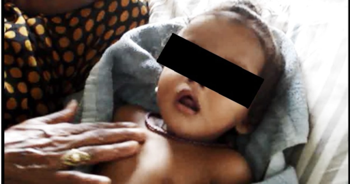
Toxic looking, irritable, anxious and restless child prefers to sit leaning forward with mouth open with drooling of saliva. Remain clinging onto the mother.He was tachypneic with RR of 56/ min and in severe respiratory distress with suprasternal, subcostal and intercostal retractions. In order to avoid agitation and further worsening of distress the pulse oximeter was attached with the help of the mother which showed Spo2 of 91% in air.Blow-by Oxygen was administered in a non-threatening manner via a mask held by the motherat a distance from the child’s face. Despite improvement in Spo2 to 97%, respiratory distress remained the same. A provisional diagnosis of upper airway obstruction with respiratory failure was made. Anticipating urgency of total airway obstruction call was sent to ENT and anesthesia departments.
He was then shifted to PICU with oxygen accompanied by intensivist. PICU staffs got the intubation and tracheostomy equipments ready. While the IV cannulation tray was readied the child started crying and suddenly became pale, desaturated and collapsed. Bag and Mask ventilation was started and IV cannula secured.The senior most intensivist intubated the child after intravenous Fentanyl with 4mm sized ET tube (one size smaller than estimated). During intubation a pale and swollen epiglottis, normal looking glottis clear with no secretions from the subglottic region was visualized. Spo2 improved to 100% and the child was put on ventilator with minimal settings.
General examination revealed 3mm sized tender cervical lymphadenitis. High grade fever(103°F) and normal BP. On auscultation bilateral air entry was equal with no added sounds were noted .CVS, abdomen and CNS examination were normal.
Based on the above findings diagnosis to be considered include
Croup,Bacterial tracheitis,supraglottitis, angio-edema, foreign body airway obstruction.
Croup: commonest cause of upper airway obstruction in children. In our case no viral prodromal symptoms like running nose was present. No classical barking cough and prominent stridor. High grade instead of low-grade fever in LTB. Child with Croup is not so toxic, there is gradual onset and progression instead of acute onset and rapid progression seen in this patient.
Bacterial Tracheitis –Insidious onset with sudden deterioration. Toxic look, characteristic brassy cough, subglottic edema with normal epiglottis. Copious thick tracheal secretion is present
Supraglottitis – Rapid onset, minimal non barking cough, toxiclook, stridor, drooling, dysphagia, muffled voice, tripod position, cherry red or pale and edematous epiglottis, and aryepiglottic folds. Has fulminant presentation.
Angio-edema – Nontoxic, history of allergy, urticarial, abdominal pain and hypotension.
History doesn’t suggest foreign body obstruction.
PROVISIONAL DIAGNOSIS
Supraglottitis
INVESTIGATIONS
Investigations were sent and IV Ceftriaxone was started
Hb – 10.2
TLC – 25000
P83 L16 E1
X-ray Chest – NAD
Ryles tube feeding was started on day 2 of admission.
Blood CS –Methicillin sensitive Staphylococcus aureus.
Swab from supraglottic area grew Staphylococcus aureus
Finally, a diagnosis of Staphylococcal supraglottitis was made.
The child became afebrile after 72 hours, air leak developed around the endotracheal tube at 48 hours. The child was successfully extubated on day three of hospitalization. Oral feeds were started on day four and was discharged home after 7 days of parental ceftriaxone therapy.
Discussion:
Majority of cases of supraglottitis is caused by Haemophilusinfluenza,however because of widespread HiB vaccination the incidence of the disease has fallen from around 4.39 to 1.55 per 100,000. The disease now occurs in true vaccine failures, including immunocompromised hosts.Other organisms causing supraglottitis include groups ß-hemolytic Streptococcus, H parainfluenzae, Staphylococcus aureus,Streptococcus pneumoniae, Klebsiella, Pseudomonas, Candida and viruses. Our case was fully vaccinated hence the disease was not caused by HiB. The disease tends to occur in children 2 to 7 years of age, but cases have been reported in those younger than 1 year of age.
Classic epiglottis is a fulminant disease in otherwise healthy child, who can be near death in a few hours.The disease has not been eliminated, but due to its rarity there are concerns about a potential lack of familiarity with its management among emergency physicians, pediatricians,ENT surgeons and anesthesiologists. Up to 20% of infants with epiglottitis are misdiagnosed initially, usually with viral LTB.
The first priority and key to the diagnosis is strong suspicion and to secure the airway in a controlled environment. The examination (especially of the throat) and cannulation or venipuncture should be deferred, because emotional upset and crying may precipitate complete airway obstruction.The child should be approached in a calm and reassuring manner.